Kidney CT Appearances
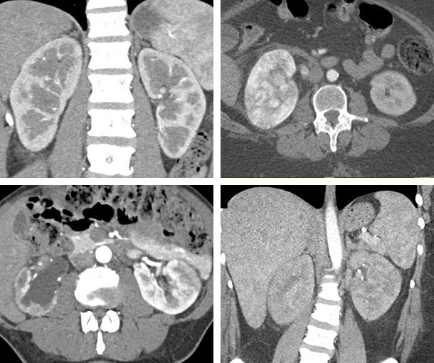
Acute Pyelonephritis CT Findings
- IV contrast needed
- Poorly defined areas of hypoattenuation in the kidney
- Can be focal, unilateral or bilateral
- Extends from the papillary to the cortical surface
- May or may not have swelling of the kidney
- Reduced corticomedullary differentiation (classic)
- May be difficult occasionally to distinguish from a renal infarct
Related Lectures:
CT Evaluation of Hematuria: A Practical Approach Part 1
CT of the Acute Abdomen: GU Applications Part 1
CT Evaluation of Hematuria: A Practical Approach - Part 1