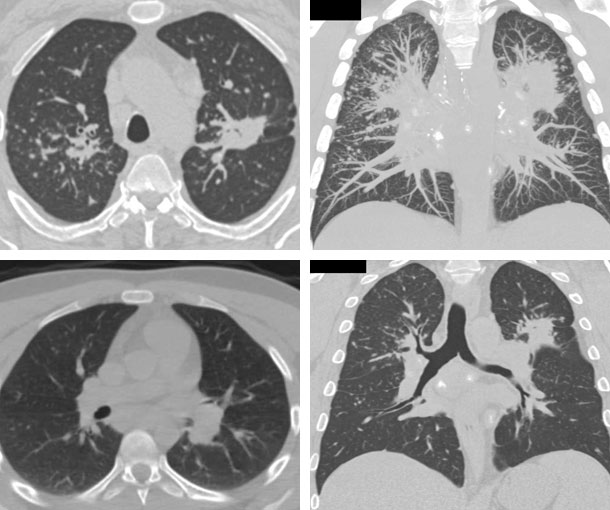
Chest CT Appearances
Sarcoidosis of the Lungs CT Findings
- Well-defined mediastinal and hilar lymph adenopathy
- “1-2-3 pattern” is combination of right paratracheal, right hilar, and left hilar lymph node enlargement
- Nodules grouped along the bronchial vascular bundles, interlobular septa, interlobar fissures, and subpleural areas
- Nodules can merge to form larger opacities
- Regional air trapping
- Stage I: presence of bilateral hilar adenopathy
- Stage II: bilateral hilar adenopathy and pulmonary infiltrates
- Stage III: pulmonary infiltrates without overt hilar adenopathy
- Stage IV: presence of overt pulmonary fibrosis
Related Pearls: Sarcoidosis